Principal Investigator:
Susan C. Pannullo, Professor of Clinical Neurological Surgery
Background & Unmet Need
- Radiosurgery, or stereotactic surgery, is a noninvasive technique in which focused radiation beams are delivered into the body for treatment of tumors or other abnormalities
- Stereotactic surgery does not require any incisions, and instead uses high resolution, 2D brain scans to target the radiation
- Targeting radiation to minimize damage to surrounding tissues requires a multidisciplinary team
- During radiosurgery case planning, team members must mentally convert complex neural anatomy from 2D brain scans to 3D abstraction
- The ability of team members to do this conversion varies, and current commercially available radiosurgery software allows simulated 3D projections only
- Unmet Need: Precise and anatomical 3D visualization of neural anatomy for surgical case planning and guidance
Technology Overview
- The Technology: A system for visualization of neural anatomy which uses augmented reality to render 3D images of scans
- Volumes of neural structures are obtained by CT/MRI and are processed to build a holographic model resembling the intracranial structures
- A computer program precisely renders the anatomy and allows the user to reconstruct regions of interest, which is projected via a Microsoft HoloLens headset
- PoC Data: Interactive models of neuro-oncologic scenarios have been created, which have demonstrated 3D identification of the following structures:Intercranial metastases,Resection cavity with metastasis,Intraventricular lesion
Technology Applications
- Enhances radiosurgery case planning discussions and allows for practicing in simulations
- Detection of tumors, lesions, and cysts on key organs
- Reconstruction of organs for root-cause analysis of medical conditions
- Use in medical education to study highly variable conditions
Technology Advantages
- Offers 3D views and cross-sectioning capabilities, whereas current imaging technologies only offer views in 2D
- Allows for real-time interaction with the patient’s model, such as moving the model around, hiding structures, fading structures, and layering with 2D imaging
- Compatible with off-the-shelf VR hardware
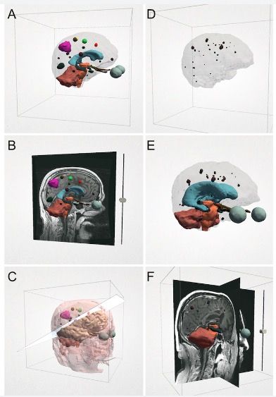
Figure 1: NeuroVis holograms for a patient undergoing stereotactic radiosurgery planning for treatment of a resection cavity with metastasis (A-C) and for a patient with multiple intracranial metastases (D-F).
Publications
Resources
Intellectual Property
Patents
- US Patent Application Filed
Cornell Reference
- 9511
Contact Information

For additional information please contact
Donna Rounds
Associate Director, Business Development and Licensing
Phone: (646) 780-8775
Email: djr296@cornell.edu